


UNH Pathologist Diagnoses Fungal Disease in Rescue Dog from Arizona
DURHAM, N.H.—A pathologist with the New Hampshire Diagnostic Veterinary Lab at the University of New Hampshire recently diagnosed the fungal disease Valley Fever in a rescue dog from Arizona. It is the first time the lab has diagnosed this disease in a dog in the state. The disease, which is treatable, is endemic in the Southwest and rarely seen in native New England dogs.
“This case serves as a reminder to inform your veterinarian of your pet’s travel history and the importance of submitting biopsied tissues for examination even if the lesion seems routine,” said Colleen Monahan, senior veterinary pathologist and clinical assistant professor. “This is especially important given the increased number of homeless dogs and cats being transported from southern states to New England for adoption. These animals may be infected with diseases that are endemic in those states but not normally seen in this region.”
Monahan made the diagnosis after a mass on the leg of a New Hampshire dog, removed by a veterinarian, was submitted to the lab for biopsy.
“There are rare reports of people and animals from New England contracting Valley Fever after travelling to a region where it is more prevalent,” she said. “In this case, we learned from the veterinarian that this dog was rescued from Arizona two years prior and had a small mass on its leg at that time. The mass recently had increased in size, leading to concern from the owner and removal by the veterinarian.”
According to the Valley Fever Center for Excellence at the University of Arizona, two-thirds of U.S. Valley Fever infections are contracted in Arizona even though nationally the disease is uncommon. People and animals can develop Valley Fever after inhaling spores growing in loose, sandy soils most commonly found in the southwestern United States and northwestern Mexico. The disease cannot be passed from animals to people or between animals.
The most common early symptoms are coughing, fever, weight loss, lack of appetite and lack of energy. Most dogs, with adequate antifungal therapy, recover from the disease, especially with early diagnosis and intervention.
“While there are tests that can be run to rule out Valley Fever, since it is so rare a diagnosis in this region, screening testing of adopted dogs would be excessive. Instead, owners should inform their veterinarians if their animals were adopted from or traveled to different regions—especially if the animal is sick—so the veterinarian can have the whole picture to better diagnose and treat their pet,” Monahan said.
As part of the NH Agricultural Experiment Station at UNH, the New Hampshire Veterinary Diagnostic Laboratory serves the state of New Hampshire as a key partner with the New Hampshire commissioner of agriculture and state veterinarian in their efforts to monitor and control important animal diseases. The lab also provides diagnostic services to veterinarians from New Hampshire and New England who use the lab’s services for the diagnosis of animal diseases in pets, farm animals, wildlife, zoo, and marine animals.
Founded in 1887, the NH Agricultural Experiment Station at the UNH College of Life Sciences and Agriculture is UNH’s original research center and an elemental component of New Hampshire's land-grant university heritage and mission.
The University of New Hampshire inspires innovation and transforms lives in our state, nation and world. More than 16,000 students from all 50 states and 71 countries engage with an award-winning faculty in top-ranked programs in business, engineering, law, health and human services, liberal arts and the sciences across more than 200 programs of study. As one of the nation’s highest-performing research universities, UNH partners with NASA, NOAA, NSF and NIH, and receives more than $110 million in competitive external funding every year to further explore and define the frontiers of land, sea and space.
PHOTO AVAILABLE FOR DOWNLOAD
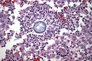
https://colsa.unh.edu/nhaes/sites/default/files/media/images/coccidioides.jpg
Photomicrograph from the biopsied mass showing a fungal spherule (arrow), consistent with Coccidioides, surrounded by inflammatory cells. Credit: NH Veterinary Diagnostic Lab
-
Media Contact
Lori Tyler Gula, PhD | NH Agricultural Experiment Station | lori.gula@unh.edu | 603-862-1452
Latest News
-
January 12, 2026
-
December 4, 2025
-
November 26, 2025
-
November 6, 2025
-
November 5, 2025